How virtual reality is transforming surgery
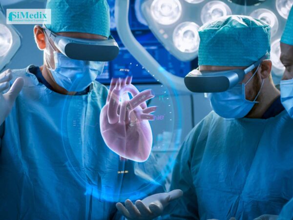
Virtual reality technology enables surgeons to virtually explore a patient’s anatomy prior to performing a procedure, much like a pilot uses a flight simulator. The technology can also be used as a teaching tool for medical students and residents.
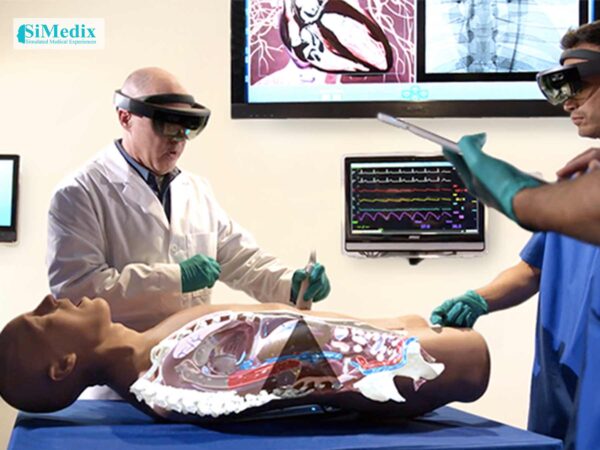
Danielle Collins had a brain bleed caused by arteriovenous malformation (AVM), an abnormal connection between the arteries and veins. She was admitted to the George Washington University Hospital, where her medical team explained everything that was happening with support from Precision Virtual Reality™ technology, an advanced system that gives patients a 360-degree view inside their own anatomy. Using a touch screen and a virtual reality headset, Danielle was able to get different views of her brain by turning her head and moving the joy stick. She says that seeing the images helped “take away the fear of the unknown,” and she was able to process her diagnosis and treatment plan on a physical and emotional level.
The technology helped her doctors, as well, by enabling them to map out alternate surgical approaches ahead of time and find the safest and best possible option. After her craniotomy procedure to remove the AVM, Danielle again used the virtual reality technology, this time to confirm that the AVM was gone. Seeing the results for herself alleviated any potential anxiety and reaffirmed that she was going to be okay. “To be able to see that is amazing,” she says.
The next frontier in virtual reality technology
Many of the possible uses for the technology are yet to be realized, says Anthony Caputy, MD, FACS, chairman of the Department of Neurosurgery at GW Hospital. Its application is already being expanded at GW Hospital to benefit patients and doctors in the treatment of thoracic conditions. Much like the brain, the chest has major blood vessels and anatomy where millimeters matter when performing advanced medical procedures. Thoracic surgeons at the hospital are now using Precision Virtual Reality technology to support surgical planning and patient education. Also, the hospital has been awarded a medical education grant to study the usefulness of VR imaging as a tool for staging lung cancer, says Keith Mortman, MD, director of thoracic surgery. Staging involves assessing the extent to which cancer has spread.
Dr. Mortman notes that the VR platform can be beneficial for treating patients who have a mass in their chest that may be next to or invading other structures. The advanced 360-degree view enables surgeons to better see where structures, vessels and airways are in relation to one another. This can help determine the best treatment and whether certain surgeries can be safely and effectively performed.
Surgery is not recommended for tumors that have spread to the middle part of the chest, between the lungs, says Dr. Mortman. VR technology can potentially help to identify if a mass has invaded that area, and thereby avoid unneeded procedures. In the event that surgery is recommended, more precise views can help direct treatment and possibly change the approach. For example, instead of going through the front of the chest, the surgeon may decide that it’s better to go through the side.
Along with the potential for improving medical care, VR capabilities also offer other advantages. Patients can put on the goggles and become an “avatar” walking through their chest, Dr. Mortman says. This can provide a new and compelling way to help patients better understand their diagnoses, and be more involved in their treatment.